Retrobulbar neuritis
- Reviewed by Robert H. Shmerling, MD, Senior Faculty Editor, Harvard Health Publishing; Editorial Advisory Board Member, Harvard Health Publishing
What is retrobulbar neuritis?
Retrobulbar neuritis is a form of optic neuritis in which the optic nerve, which is at the back of the eye, becomes inflamed. The inflamed area is between the back of the eye and the brain. The optic nerve contains fibers that carry visual information from the nerve cells in the retina to the nerve cells in the brain. When these fibers become inflamed, visual signaling to the brain becomes disrupted and vision is impaired.
Retrobulbar neuritis can be caused by a variety of conditions, including:
- infections such as meningitis, syphilis, and various viral illnesses
- multiple sclerosis
- tumors
- exposure to certain chemicals or drugs
- allergic reactions.
However, in many cases the cause is unknown. Vision loss can be minimal, or the disease can result in complete blindness.
Optic neuritis affects women twice as often as men, and usually affects adults between the ages of 20 and 40.
The vast majority also have pain when they move their eyes. Retrobulbar neuritis often is an early sign that someone has multiple sclerosis. Between 20% and 40% of the 20,000 people who develop optic neuritis in the United States each year will develop multiple sclerosis within five years.
Symptoms of retrobulbar neuritis
Symptoms usually worsen for two weeks and then stabilize. However, the course of the illness varies greatly. Most cases show improvement over time, although persistent visual impairment is common. Optic neuritis usually affects only one eye, but both eyes may be affected. Common symptoms include:
- blurred or dimmed vision
- a blind spot at or near the center of vision
- color washout so that colors are less rich
- pain with eye movement
- tenderness of the eye to touch or pressure
- complete blindness in the affected eye.
Diagnosing retrobulbar neuritis
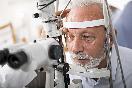
A doctor will use an ophthalmoscope or other specialized equipment (such as a slit lamp) to examine the back of the eye, particularly the optic disc. This is where the optic nerve fibers concentrate before exiting the eye to extend back toward the brain. In the early stages of retrobulbar neuritis, the optic disk may appear normal. Later, it may become pale.
The pupil normally becomes smaller (constricts) in response to light. In retrobulbar neuritis, this response often is reduced in the affected eye. The doctor also will test your visual acuity, which frequently is impaired in the affected eye. The doctor will test your side (peripheral) vision because, in many cases of retrobulbar neuritis, a scotoma (a blind or dark spot in the visual field) may be detected. The doctor also may search for associated conditions such as infection or multiple sclerosis, after a detailed discussion about other symptoms and a complete physical examination.
Expected duration of retrobulbar neuritis
How long this condition lasts depends on the cause, and in some people optic neuritis recurs or becomes chronic. If the optic nerve is permanently damaged, it can lead to blindness.
Preventing retrobulbar neuritis
Because the underlying cause of most cases of retrobulbar neuritis is unknown, there is usually no way to prevent it. Practicing safe sex to avoid certain infections such as syphilis and being cautious around chemicals and toxins is always wise.
Treating retrobulbar neuritis
Many cases improve without treatment. A corticosteroid medication, such as intravenous methylprednisolone, may be recommended, especially if vision loss is significant or if an MRI shows abnormalities suggestive of multiple sclerosis. Additional treatments for suspected multiple sclerosis may be offered, including interferon-beta, glatiramer acetate (Copaxone), dimethyl fumarate (Tecfidera), or teriflunomide (Aubagio); these medications may reduce the chances of recurrent neuritis or other symptoms of multiple sclerosis.
When to call a professional
Call a doctor if you experience any vision changes, either suddenly or over time. Pain in or around the eye, with or without vision loss, also should receive prompt medical attention.
Prognosis
The outlook depends on the cause. Cases in which there is no obvious cause, or in which the cause is multiple sclerosis, often improve after two weeks, but the vision may never completely return to normal.
Retrobulbar neuritis may return, and many people with retrobulbar neuritis eventually develop multiple sclerosis. If an MRI image of the brain is abnormal in a manner typical of multiple sclerosis at the time of retrobulbar neuritis, clinically obvious multiple sclerosis is much more likely than if the MRI is normal.
Additional info
National Eye Institute
http://www.nei.nih.gov/
American Academy of Ophthalmology
http://www.aao.org/
About the Reviewer

Robert H. Shmerling, MD, Senior Faculty Editor, Harvard Health Publishing; Editorial Advisory Board Member, Harvard Health Publishing
Disclaimer:
As a service to our readers, Harvard Health Publishing provides access to our library of archived content. Please note the date of last review or update on all articles.
No content on this site, regardless of date, should ever be used as a substitute for direct medical advice from your doctor or other qualified clinician.


