


Surgery for a retinal detachment
Occasionally, the vitreous gel (the egg white–like substance that fills most of the eyeball) pulls on the retina with enough force to tear it. This separation of the retina from the back of the eye allows fluid from inside the eye to enter through this tear and detach the retina from the choroid (the nutrient-rich layer underlying the retina). If this rupture is caught and treated early, a retinal detachment (see illustration) may be prevented.
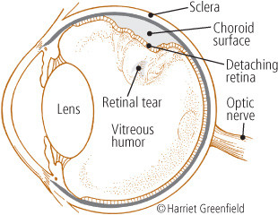
Retinal detachment
If not treated, the retinal detachment may continue until the retina is nearly totally detached from the back of the eye and maintains a connection only at the optic nerve in the back of the eye and the ciliary body (a ring of tissue that encircles the lens) in the front of the eye.
To continue reading this article, you must log in.
Subscribe to Harvard Health Online for immediate access to health news and information from Harvard Medical School.
- Research health conditions
- Check your symptoms
- Prepare for a doctor's visit or test
- Find the best treatments and procedures for you
- Explore options for better nutrition and exercise
I'd like to receive access to Harvard Health Online for only $4.99 a month.
Sign Me UpAlready a member? Login ».
Disclaimer:
As a service to our readers, Harvard Health Publishing provides access to our library of archived content. Please note the date of last review or update on all articles.
No content on this site, regardless of date, should ever be used as a substitute for direct medical advice from your doctor or other qualified clinician.